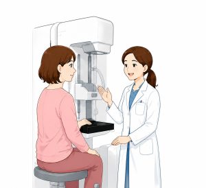

Differences from traditional mammograms and benefits for Japanese women
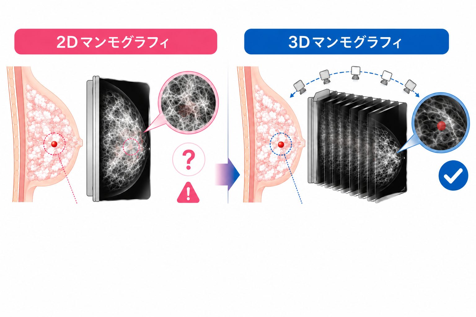
Are you familiar with the 3D mammography (tomosynthesis) imaging method? Conventional mammography presses down on the breast and takes a two-dimensional (2D) image from both directions. With 3D mammography, the X-ray tube rotates while maintaining the same pressure, taking multiple images, and reconstructing the interior of the breast as thin slice images (sectional views).
In other words, the biggest difference between conventional mammography and 3D mammography is that the former is "planar," while the latter can be "observed in three dimensions."
Breasts are structured with overlapping fat and mammary glands. Therefore, conventional 2D mammography can hide small lesions caused by overlapping glands, or conversely, normal mammary glands can appear “solid.” This can lead to the need for a more detailed examination even when there is no actual disease.
On the other hand, 3D mammography allows for the internal structure of the breasts to be seen through a cross-sectional view, allowing for the separation and confirmation of breast tissue abnormalities. As a result, even small tumors that were previously difficult to see and structural abnormalities can now be detected more accurately. As a result, it is expected to reduce unnecessary re-testing while increasing the detection rate of early breast cancer.
Furthermore, Japanese women tend to have a higher concentration of dense breasts (densest breasts) compared to Western women. In this case, conventional 2D mammography results in white images of the breasts and tumors, which has the disadvantage that the lesions are difficult to detect in the background. However, 3D mammography allows for the observation of breast overlap on the cross-section, reducing the risk of missed findings in cases of densest breasts, making it a beneficial test.
In recent years, 3D mammography has been introduced not only in universities and large facilities but also in local clinics, and it is sometimes included as a part of regular health check-ups. The availability of high-precision testing at local medical institutions leads to the creation of a healthcare environment that makes it easier for patients to seek medical care.
Furthermore, pregnant women or those who may be pregnant are not eligible to undergo X-ray mammography for breast cancer screening. In that case, we recommend performing a breast ultrasound examination. Receiving a proper and highly accurate examination is an important step towards early detection of breast cancer.
Point

3D mammography allows for the visualization of the breasts in three dimensions as a cross-sectional image, allowing the separation and confirmation of breast tissue overlap.
Information

Japan Green Vietnam Clinic
Japan Green Vietnam Clinic
Mitchiko Yanano
After obtaining a radiology technician license in Japan, I gained experience working in hospitals and clinics. Subsequently, I worked at the Shanghai Green Clinic of the JGH Group for seven and a half years. Starting with 3D mammography, we will continue to support Vietnam in providing accurate and reliable examinations using the latest X-ray equipment.
AddressLac Hong Lotus Building 1-N01-T5, Hoang Minh Thao, Xuan Dinh, Hanoi
Phone number: 024-7300-0388 (Japanese available)
Consultation hours: Monday to Friday 9:00 AM to 12:00 PM, 1:00 PM to 5:00 PM, Saturday 9:00 AM to 12:00 PM *Closed on Sundays
website https://www.japangreen.vn/
Instagram japangreen.vn